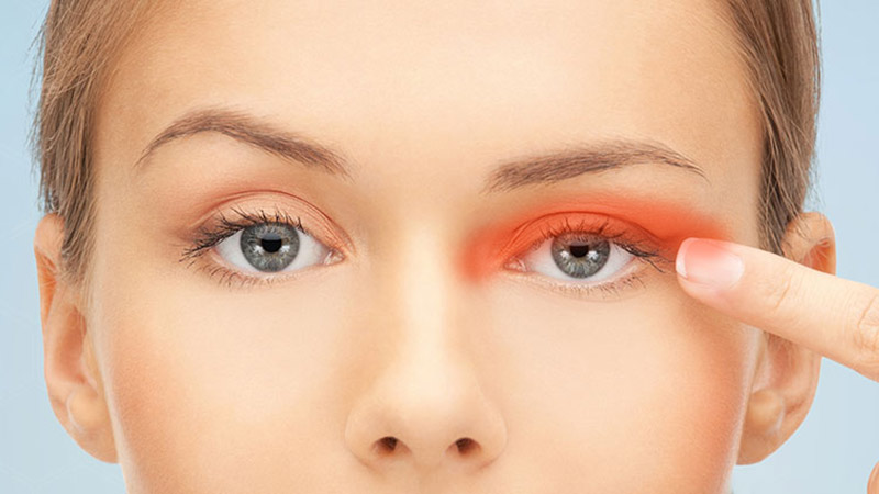
A xanthelasma is a benign growth on the eyelid skin caused by a local build-up of lipoproteins in the inner layer of the skin (dermis). It is usually located ...
Price from 400 €Price on 10.09.2026.: 400 €A xanthelasma is a benign growth on the eyelid skin caused by a local build-up of lipoproteins in the inner layer of the skin (dermis). It is usually located in the skin of the inner corner of the upper and lower eyelid in the form of yellowish-white, sharply defined plaques, and it spoils the aesthetic appearance of the face. They are removed surgically.
Xanthelasmas in themselves pose no danger to the eye, but they can point to a possible disorder of liver and pancreas function, so further investigation is recommended.
They most often appear as yellowish-white, sharply defined plaques located in the inner corner of the eyelids. Xanthelasmas are soft to the touch and can appear on all four eyelids. Over time they can also grow and merge with each other. They do not interfere with eyelid function unless ptosis (a drooping upper eyelid) develops.
They most often develop because of raised blood fat levels, but can also occur with diabetes, liver disease and genetic factors.
Incidence in men/women, age group
It occurs more often in post-menopausal women.
In fact, there is no natural treatment; they always need to be removed surgically. As a precaution, a blood test should be done to establish whether the patient has raised blood fats, and the patient should be advised to get them under control to reduce the chance of the xanthelasmas returning in future.
A detailed specialist examination with a slit lamp (biomicroscope) is needed before xanthelasma surgery.
The patient is told what the procedure will look like, then receives the necessary premedication.
On entering the operating theatre the patient is given a sterile cap and shoe covers.
In the operating theatre the patient is made comfortable on a soft bed. Nurses disinfect the skin around the eyes and place a sterile drape over the patient.
The procedure takes 15-30 minutes, depending on the size and number of growths to be removed. The patient lies on the bed and is awake and in communication with the surgeon throughout. The skin is anaesthetised, so the procedure is completely painless and comfortable. Because the operation is elegant and quick, most patients are surprised at how fast and simple everything was and that all the fear people usually have before a procedure was unfounded.
After the operation a sterile pad is placed over the eye. On discharge from the clinic the patient receives post-operative treatment consisting of drops and/or ointments, along with instructions to follow. Three hours after the procedure the patient removes the pad and starts the prescribed treatment. The patient returns for a check-up the next day, and until then should avoid unfavourable environmental conditions (wind, dust, smoky rooms). One more check-up is needed after 7-10 days to remove the stitches (if there are any), and then as needed.
is an outward turning of the eyelid, so that the conjunctiva faces outwards and is exposed to external influences, which can cause dryness, redness and a gritty feeling in the eye. If it affects the lower eyelid it can prevent tears from draining, so constantly wiping them away makes the condition even worse.
It is more often present on the lower eyelid and can be congenital or acquired. Congenital ectropion is rare and is most often associated with other anomalies. Acquired ectropion can be:
Involutional ectropion results from a loss of tension in the tarsus and eyelid ligaments due to ageing.
Mechanical ectropion is caused by a growth that mechanically turns the eyelid out.
Cicatricial ectropion is the result of scarring of the skin after injury or inflammation.
Paralytic ectropion occurs after paresis of the facial nerve (n. facialis), which leads to loss of function of the orbicularis oculi muscle and the inability to close the eye (lagophthalmos).
Neutral and antibiotic drops and ointments are used to relieve symptoms temporarily, while a definitive cure is achieved with an operation to shorten the eyelid.
is an inward turning of the eyelid margin towards the eyeball, so that the eyelashes come into contact with the cornea and conjunctiva, which gives a feeling of a foreign body in the eye and scratching.
Congenital entropion results from an underdeveloped or insufficient tarsus and a hypertrophic marginal part of the orbicularis muscle, which leads to a loss of eyelid stability. Acquired entropion can be:
Involutional entropion most often appears on the lower eyelid after the age of 60. It results from a loosening of tension in the eyelid structures due to ageing.
Mechanical entropion usually appears on the upper eyelid due to mechanical pressure from excess skin, tissue swelling or a tumour near the eyelid.
Cicatricial entropion results from scarring changes of the eyelid due to inflammation, injury or surgery.
Spastic entropion is caused by a reflex spasm of the orbicularis oculi muscle due to irritation or inflammation of the eye.
Neutral and antibiotic drops and ointments are used to relieve symptoms temporarily, while a definitive cure is achieved with an operation on the eyelid.
Točan iznos ovisi o nalazu i procjeni liječnika. Sve cijene pogledajte u cjeniku.
Full price listCijena prema važećem cjeniku poliklinike.
Specialist in ophthalmology, founder of Poliklinika Knezović
Expert in diseases of the anterior segment of the eye and in laser treatment of keratoconus. More than 25 years in ophthalmology, with training at leading centres around the world.
Every eye is different. The doctor decides on the method only after an examination and diagnostics. Book an appointment and you will receive a clear recommendation and an exact quote, with no obligation.