
Retinal detachment is an emergency in which the retina (nerve tissue at the back of the eye) separates from its underlying layer, thus losing its nourishment. The retina...
Price of the procedure 1.330 €Price on 10.09.2026.: 1.330 €Retinal detachment is an emergency in which the retina (nerve tissue at the back of the eye) separates from its underlying layer, thus losing its nourishment. The retina is nerve tissue that converts light stimuli into an electrical signal, which travels along the optic nerve to the brain, where we experience the image.
This type of retinal detachment is the most common. It is caused by a hole or tear in the retina that allows fluid to pass through and collect beneath the retina, separating it from its underlying layer. The detached area is thus left without nourishment and loses function, resulting in loss of vision.
The most common cause of rhegmatogenous detachment is ageing. With age the vitreous (the gel-like substance that fills the eye and gives it volume) changes its properties and becomes thinner. Usually the vitreous separates from the surface of the retina without any complications – a phenomenon called posterior vitreous detachment (PVD). One of the complications of such detachment is a retinal tear.
This form occurs when scar tissue grows on the surface of the retina, pulling the retina and separating it from its underlying layer. It usually occurs in people with poorly controlled diabetes or similar conditions.
In this form fluid collects beneath the retina, but there are no holes or tears in the retina. Exudative detachment can be caused by age-related macular degeneration, eye injury, a tumour or inflammatory disorders.
The following factors increase the risk of retinal detachment:
Retinal detachment is painless, but certain warning signs are almost always present:
A complete ophthalmological examination in which the ophthalmologist examines your eye in detail. The pupils need to be dilated so that the examination of the back of the eye (fundus) is better and the periphery of the fundus can also be examined. Other tests may also be indicated at the examination, such as an eye ultrasound or OCT.
Except in the earliest stage of the disease, when any retinal defect can be repaired with laser “seals” around the lesion, surgical treatment is almost always needed to repair the hole, tear or detachment of the retina. There are several different surgical techniques.
When a retinal hole has not yet progressed to detachment, the ophthalmologist may suggest one of the following procedures to prevent retinal detachment and preserve vision.
The surgeon directs a laser beam into the eye through the pupil. The laser creates “seals” around the retinal defect, forming scars that usually “weld” the retina to the underlying tissue.
After a local anaesthetic is applied, the surgeon applies a freezing probe to the outer surface of the eye directly at the site of the defect. The freezing creates a scar that helps secure the retina to the wall of the eye.
Both procedures are performed on an outpatient basis and it is necessary to avoid physical activity for a while – such as running – for a couple of weeks or more.
If the retina has completely detached – surgery is needed to repair the condition, preferably within a few days of diagnosis. The type of surgery the vitreoretinal surgeon recommends will depend on several factors, primarily the severity of the detachment.
In this procedure, called pneumatic retinopexy, the vitreoretinal surgeon injects a bubble of air or gas into the central part of the eye. Properly positioned, the bubble pushes the area of the retina with the defect against the wall of the eye, stopping further passage of fluid into the subretinal space. The surgeon then uses cryopexy (freezing) to repair the retinal defect.
The fluid that has collected beneath the retina is absorbed on its own, and the retina can then return to its natural position, against the wall of the eye. After the operation the patient usually has to keep the head in a certain position for up to several days so that the bubble stays in the required position. The bubble is reabsorbed after a few days.
In this procedure the surgeon places a silicone buckle over the affected area, cancelling out the force with which the vitreous pulls on the retina.
If the patient has several holes or defects, the surgeon may place a scleral band that encircles the whole eye like a belt. The band is positioned so that it does not interfere with vision and usually remains permanently.
In this procedure the surgeon removes the vitreous together with any tissue pulling on the retina. Air, gas or silicone oil is then injected in place of the vitreous to flatten the retina.
Eventually the air, gas or fluid is reabsorbed and the vitreous space refills with the body's own fluid. If silicone oil is used, it can be surgically removed months later.
Vitrectomy can be combined with placement of a silicone buckle.
After the operation it can take several months for vision to improve. Sometimes a second operation is needed for successful treatment. The success of the operation depends on how well the retinal tissue has survived the interruption of blood supply and nourishment.
Before the operation
Since time, along with the severity of the detachment itself, is a key factor, it is important to come for an eye examination as soon as possible after symptoms appear.
How the operation goes
Eye surgery is performed under local anaesthesia with sedation. This means the patient receives a local anaesthetic next to the eye and sedatives. The operation itself is painless and lasts on average from an hour to several hours. After the operation the patient goes home.
After the operation
Further follow-up examinations are scheduled the day after the examination. The eye is usually red for a few days after the operation, and the patient applies the prescribed drops and ointment.
Poliklinika Knezović has many years of experience with various modalities in the monitoring and treatment of retinal detachment and a top team of experts working in this field. We were among the first in the region to introduce layered imaging of the back of the eye (SOCT) as a prerequisite for diagnosis and treatment monitoring in retinal detachment. We are equipped with top-quality lasers for retinal photocoagulation as well as modern vitrectomy machines for performing these demanding operations. To date, several thousand procedures have been performed on the posterior segment of the eye, with excellent results.
Retinal detachment - PRICES are listed in the clinic's current price list.
Prim.dr.sc Borna Šarić is among the top vitreoretinal surgeons, with many years of experience in the diagnosis and treatment of diseases of the posterior segment of the eye. He is a regular lecturer at national and international professional conferences and a pioneer in performing the most complex operations on the anterior and posterior segments of the eye.
The prognosis of the disease and the quality of vision after surgery depend greatly on the severity of the disease, the location of the retinal detachment and how soon you see your ophthalmologist.
If untreated, retinal detachment usually spreads and affects a larger part of the eye. As a result, visual acuity is lost, which is difficult to repair if a longer period of time passes.
Točan iznos ovisi o nalazu i procjeni liječnika. Sve cijene pogledajte u cjeniku.
Full price listCijena prema važećem cjeniku poliklinike.
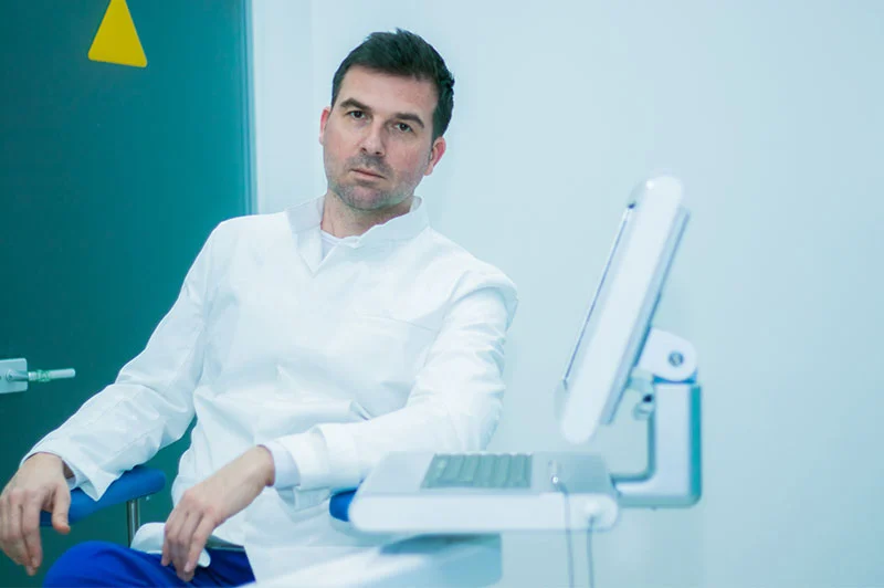
Specialist in ophthalmology, founder of Poliklinika Knezović
Expert in diseases of the anterior segment of the eye and in laser treatment of keratoconus. More than 25 years in ophthalmology, with training at leading centres around the world.
Every eye is different. The doctor decides on the method only after an examination and diagnostics. Book an appointment and you will receive a clear recommendation and an exact quote, with no obligation.