OCT
By using OCT it's possible to detect several retinal and optic nerve diseases at earliest stage.
Book an appointment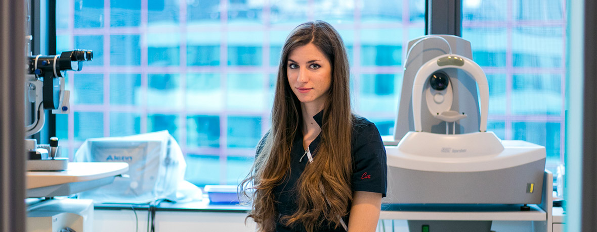
By using OCT it's possible to detect several retinal and optic nerve diseases at earliest stage.
Book an appointment
By using OCT it’s possible to detect several retinal and optic nerve diseases at earliest stage. We especially emphasize early glaucoma diagnostics, where measuring thickness of nerve fibers around retinal nerve fiber layer (RNFL) and analyzing the head of optic nerve (PNO) can discover earliest stage of glaucoma disease that is impossible to detect with other methods.
Except for early detection, this diagnostic method has important role in treatment planning (both laser and surgical), i.e. tracking different retinal and optic nerve diseases.
Optical Coherence Tomography (OCT) is new, simple, non-invasive, contactless method of laser layered scan of retina and optic nerve.
Newest Copernicus SOCT software allows detailed analysis of anterior segment of eye condition with 3-micron resolution.
By using special contactless magnifier that is adjusted do SOCT Copernicus device it’s possible to scan anterior segment of eye by this non-invasive method within only few seconds.
Scanning allows:
By using OCT it’s possible to detect several diseases at earliest stage.
Except for early detection, this diagnostic method has important role in treatment planning (laser diopter removal, anterior and posterior segment of eye operations), i.e. tracking different corneal and optic nerve diseases.
For all the questions fell free
to call us 01 / 66 77 222

Poliklinika Knezović
Green Gold Business Center, Tower V1, 8th floor
Ulica grada Vukovara 269f
10000 Zagreb
How to get to us?